
Intraoral Cameras
Intraoral Cameras
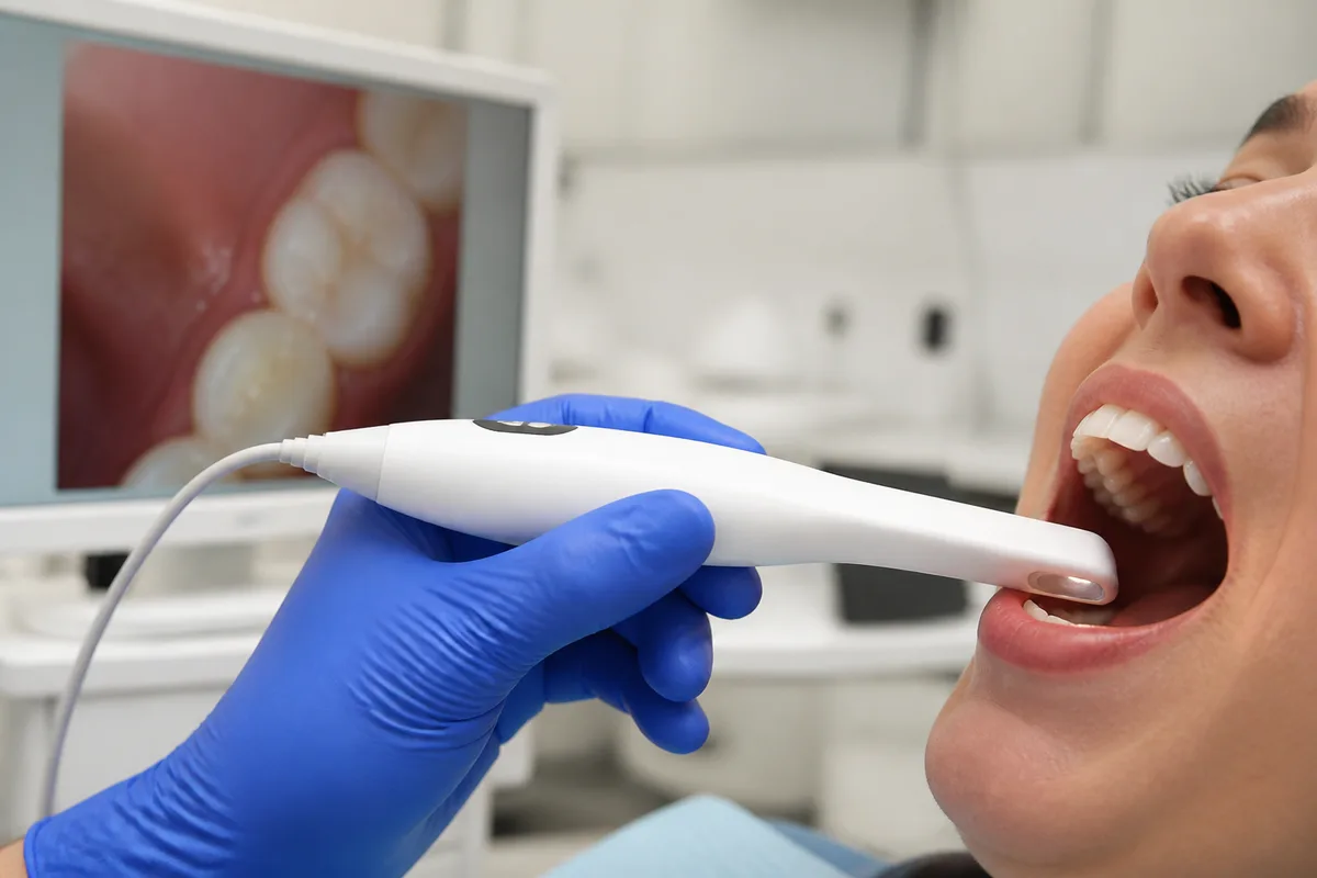
An intraoral camera is a small, pen-shaped camera that lets a dental team look closely at your teeth and gums and show you the same view on a screen. If you have ever wished you could see what your dentist sees, this is the tool that makes it possible.
What intraoral cameras are
An intraoral camera is a handheld imaging device about the size of a thick pen or a dental mirror. It has a tiny lens and a light at the tip. The tip is gently guided around the mouth, and the camera sends live images to a nearby monitor. Many cameras can also capture still pictures, so a particular tooth or area can be looked at again later.
Before cameras like this, dentists relied mostly on a small mirror, a good light, and their own eyes. Patients had to take the dentist's word for what was happening in the back of the mouth, because they could not see it. The camera improves on this in two ways. It magnifies small details, such as a hairline crack, a worn edge, or an old filling that has started to break down. It also lets the patient look along with the dentist.
The camera does not replace other diagnostic tools. It shows the surface of the teeth and the soft tissues. Images of what lies beneath the surface, such as the roots and the bone, come from X-rays.
How we use it at Brilliant Dental Group
Our facts on file for this page are limited, so we will stay general. Ask our team how the cameras fit into your particular visit. We can tell you that our comprehensive dental examination includes digital X-rays, which let us see what is going on inside your teeth and gums without cutting them open to look. A camera and X-rays answer different questions. The camera shows what is visible on the surface, and X-rays show what is hidden.
In dental practices generally, intraoral camera images are useful for several services we offer:
- Checkups and exams — a close look at tooth surfaces, gums, and existing dental work.
- Fillings and crowns — showing the wear, chips, or cracks that may call for repair.
- Periodontal care — helping patients see the condition of their gum tissue.
- Cosmetic planning — documenting the current state of your smile before whitening, bonding, or veneers.
Education is central to how Dr. Sami Hersel approaches care. He emphasizes teaching patients so they can prevent dental disease. A picture on a screen is one of the simplest ways to support that kind of conversation.
What it means for you
The biggest benefit is understanding. A problem you can see is much easier to follow than one described only in words. When a dentist points to a cracked cusp or a stained area on a screen, you can tell why a treatment is being suggested and ask better questions about it.
There are other general advantages as well:
- Clearer communication — everyone looks at the same image, which reduces guesswork.
- Early awareness — small changes are easier to notice when magnified.
- Better records — saved images can be compared over time to see whether an area has changed.
- No radiation — the camera uses visible light rather than X-rays.
- Comfort — the camera is small and is used without needles or drills.
What to expect during your visit
Before. You do not need to prepare anything special for a camera. Brush and floss as you normally would, and mention any area that concerns you, such as a sore spot, a sharp edge, or a tooth that feels different.
During. You will be seated and reclined as for any exam. The camera is moved slowly around your mouth, usually one area at a time. You may be asked to open wide or turn your head slightly. The images appear on a screen, and your dental team can explain what they see while you follow along. Most people feel only the light touch of the tip, and some notice that the lens may be briefly warm or that the cheek is gently held back. Taking pictures generally adds little time to an exam. Ask our team what to expect for your appointment.
After. There is no recovery and nothing to avoid afterward. If the images show something that needs attention, your dentist can go over the options with you while the pictures are still fresh.
Frequently asked questions
Is an intraoral camera safe?
Yes. It uses ordinary visible light and does not emit radiation. The tip is cleaned or covered between patients according to standard infection-control practice. It is a viewing tool and does not alter your teeth in any way.
Does it hurt?
It should not. The camera is small and is simply held near the teeth and gums. You might feel gentle pressure from the tip or from a cheek retractor. If any area is sore, tell your dental team so they can be extra careful there.
How often is it used?
That depends on your needs and your dentist's judgment. Cameras are commonly used at exams and when a specific concern needs a closer look or a record. Ask our team how and when they are used during your visits.
Who is it suitable for?
Intraoral cameras are generally suitable for children, adults, and older patients, since nothing is injected and no radiation is involved. They can be especially helpful for people who want to understand their treatment before deciding. If you have a strong gag reflex or a sensitive mouth, let us know and we will work with you.
If you would like to see your own smile up close or have questions about intraoral cameras, contact Brilliant Dental Group to schedule a visit.
This content is for general information only and isn't dental or medical advice. Talk with your dentist or orthodontist about your own situation. If you have a medical emergency, call 911.